-

What is DNA, and how does it determine our physical characteristics? -
To answer this question, first we must learn how DNA is structured.
Introduction to DNA
We may resemble our parents, but we are never exactly like them. This is because each child gets only some of the DNA each parent carries. About half our DNA comes from our mother, and half comes from our father. Which pieces we get is basically random, and each child gets a different subset of the parents' DNA. Thus, siblings may have the same parents, but they usually do not have exactly the same DNA (except for identical twins).
 |
|

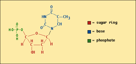
The four nucleotides look a little bit alike. They all have a ring of carbons called, in chemist's terminology, a 'sugar' (not the same as 'table sugar', however). Each nucleotide also has another type of ring structure, and this is where the four types of nucleotide are different. These rings are organic bases, much like the more familiar mineral acids and bases like NaOH or HCl, except these bases are composed of carbon, nitrogen and oxygen.
I'll try to use the term 'base' or 'basic group' to refer to just the nitrogen/carbon rings, and the term 'nucleotides' to refer to the entire structure.
Now ordinarily the atoms in a nucleotide form a three-dimensional structure. To help you visualize the structure, here's what a 'T' (T stands for Thymidine) would look like if flattened onto paper:

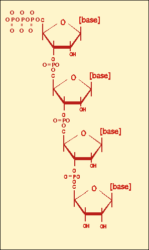
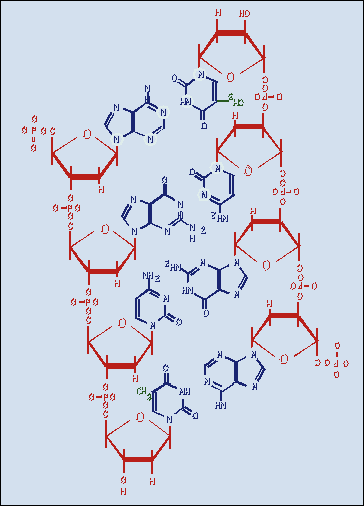
| DNA chains are made by connecting those nucleotides together via chemical bonds. At right is a diagram showing four nucleotides connected to form an oligonucleotide, in this case an RNA oligo (note that it has '-OH' at the lower right corner of each nucleotide, as opposed to the '-H' in DNA). I've left off the bases, for simplicity's sake. You can see the sugar rings linked together with phosphate bridges. This is a "single-stranded" nucleic acid. Below is the double-stranded form: |  |
 |
Double-stranded DNA is simply two chains of single-
stranded DNA, positioned so their "bases" can interact with each other. At left is a cartoon
depiction of double-stranded DNA. The sugar-and-phosphate 'backbone' is depicted in
red, and the bases are depicted in blue.
Importantly, the two strands travel in opposite directions; hence the structure is said to be "anti-parallel". The bases in the middle "pair up" with bases on the opposite strand, so that a type 'A' nucleotide is always opposite a type 'T', and 'G' is opposite 'C'. The attraction between the paired nucleotides is fairly weak, but when there is a whole string of them, it adds up to enough strength to hold the strands together. One more thing holds the strands together - an interaction called "base stacking". We don't need to consider it here. |

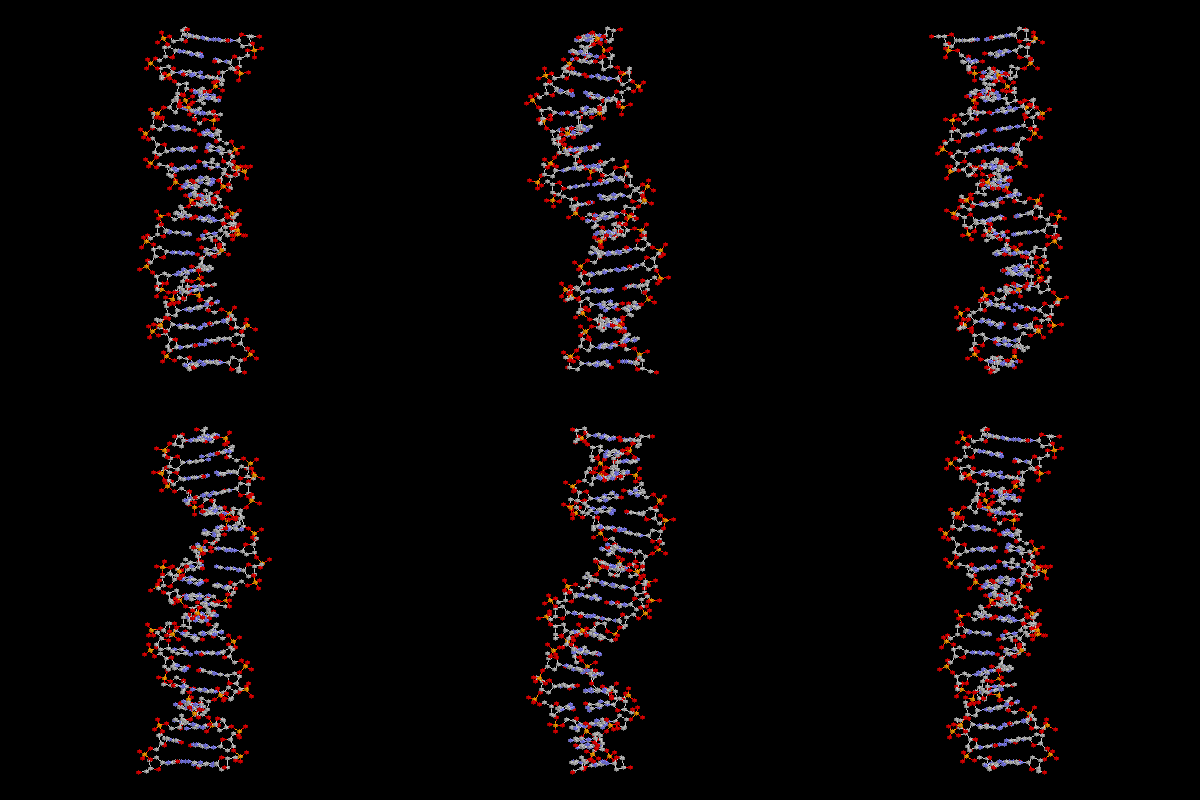
This figure was created using 'RasMol V2.6' for the Macintosh (thanks to Roger Sayle, BioMolecular Structures Group Glaxo Research & Development Greenford, Middlesex, UK.) The file 3cro.pdb was modified to remove the protein components, then a series of rotating views was screen-captured to construct this moving gif. |
At right is an animated drawing of a DNA molecule. There are two strands of DNA in this picture, wound around each other to form the famous "double helix". |
| Down each edge is the backbone, where the nucleotides are linked together to form the chain. In this drawing, you can spot the backbone most easily by looking for the red balls, the oxygen atoms. Also look for the phosphorus atoms in the backbone. These are colored yellow. Together the phosphorus and oxygen atoms form the phosphate groups that interlink the nucleotides, as described earlier. You may be able to see the sugar rings just inside the phosphate groups, visible as grey atoms - carbon. (Note that hydrogen atoms are purposely omitted from this drawing in order to simplify it). | |
| Look for the rungs of the 'ladder'. These are the basic groups that point inward and hold the two chains in position against each other. They are most easily spotted by looking for the blue color of the nitrogen atoms, which are alternating with the carbon atoms of the base groups. |