GVHD

Detection and monitoring of pulmonary complications in transplant recipients with GVHD.


Parametric Response Mapping as an Indicator of Bronchiolitis Obliterans Syndrome after Hematopoietic Stem Cell Transplantation


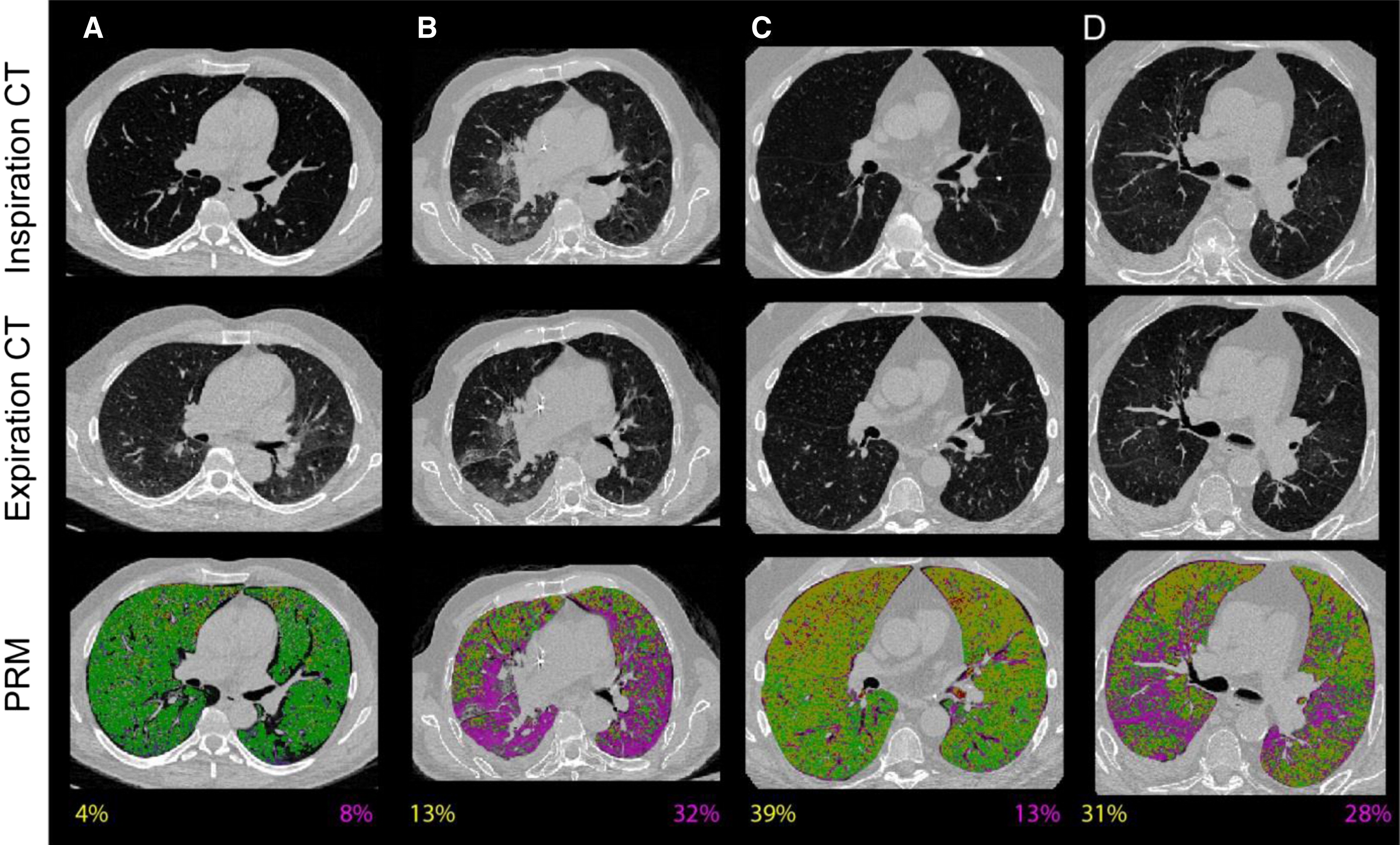
The management of bronchiolitis obliterans syndrome (BOS) after hematopoietic cell transplantation presents many challenges, both diagnostically and therapeutically. We developed a computed tomography (CT) voxel-wise methodology termed parametric response mapping (PRM) that quantifies normal parenchyma, functional small airway disease (PRMfSAD), emphysema, and parenchymal disease as relative lung volumes. We now investigate the use of PRM as an imaging biomarker in the diagnosis of BOS. PRM was applied to CT data from 4 patient cohorts: acute infection (n = 11), BOS at onset (n = 34), BOS plus infection (n = 9), and age-matched, nontransplant control subjects (n = 23). Pulmonary function tests and bronchoalveolar lavage were used for group classification. Mean values for PRMfSAD were significantly greater in patients with BOS (38% ± 2%) when compared with those with infection alone (17% ± 4%, P < .0001) and age-matched control subjects (8.4% ± 1%, P < .0001). Patients with BOS had similar PRMfSAD profiles, whether a concurrent infection was present or not. An optimal cut-point for PRMfSAD of 28% of the total lung volume was identified, with values >28% highly indicative of BOS occurrence. PRM may provide a major advance in our ability to identify the small airway obstruction that characterizes BOS, even in the presence of concurrent infection.

Read Our Paper
Research Icon


Parametric Response Mapping of Bronchiolitis Obliterans Syndrome Progression After Lung Transplantation


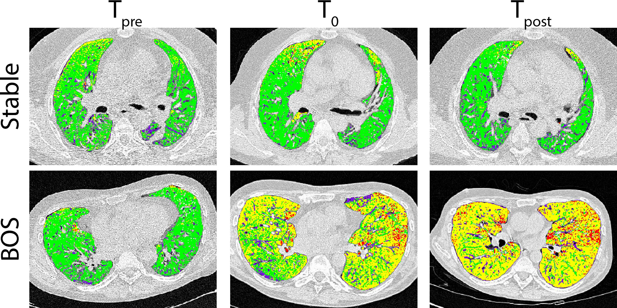
Bronchiolitis obliterans syndrome (BOS) remains a major complication after lung transplantation. Air trapping and mosaic attenuation are typical radiological features of BOS; however, quantitative evaluation remains troublesome. We evaluated parametric response mapping (PRM, voxel‐to‐voxel comparison of inspiratory and expiratory computed tomography [CT] scans) in lung transplant recipients diagnosed with BOS (n = 20) and time‐matched stable lung transplant recipients (n = 20). Serial PRM measurements were performed prediagnosis, at time of BOS diagnosis, and postdiagnosis (Tpre, T0, and Tpost, respectively), or at a postoperatively matched time in stable patients. PRM results were correlated with pulmonary function and confirmed by microCT analysis of end‐stage explanted lung tissue. Using PRM, we observed an increase in functional small airway disease (fSAD), from Tpre to T0 (p = 0.006) and a concurrent decrease in healthy parenchyma (p = 0.02) in the BOS group. This change in PRM continued to Tpost, which was significantly different compared to the stable patients (p = 0.0002). At BOS diagnosis, the increase in fSAD was strongly associated with a decrease in forced expiratory volume in 1 s (p = 0.011). Micro‐CT confirmed the presence of airway obliteration in a sample of a BOS patient identified with 67% fSAD by PRM. We demonstrated the use of PRM as an adequate output to monitor BOS progression in lung transplant recipients.

Read Our Paper

Research Icon


Parametric Response Mapping as an Imaging Biomarker in Lung Transplant Recipients


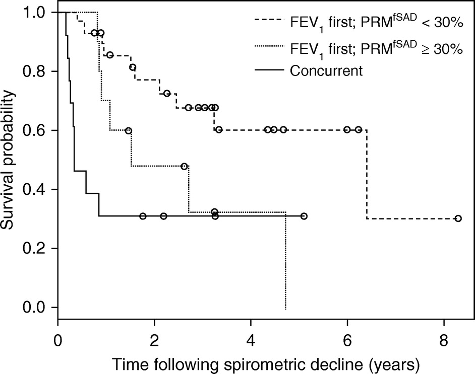
Bronchiolitis obliterans syndrome is diagnosed by spirometry, and clinical tools are lacking to identify the degree of small airway obstruction and gauge prognosis for individual patients. Recent data suggest that lung allograft rejection has multiple pathophysiologic origins with different clinical phenotypes. Quantification of functional small airways disease by parametric response mapping, a novel computed tomography–based biomarker, in lung transplant recipients with spirometric decline offers pathophysiologic information of chronic rejection phenotype. Functional small airways disease levels greater than or equal to 30% as measured by parametric response mapping identify patients with significantly shorter survival.

Read Our Paper

Research Icon


Neutrophilic Inflammation Main Culprit for Small Airway Disease Post Lung Transplant?


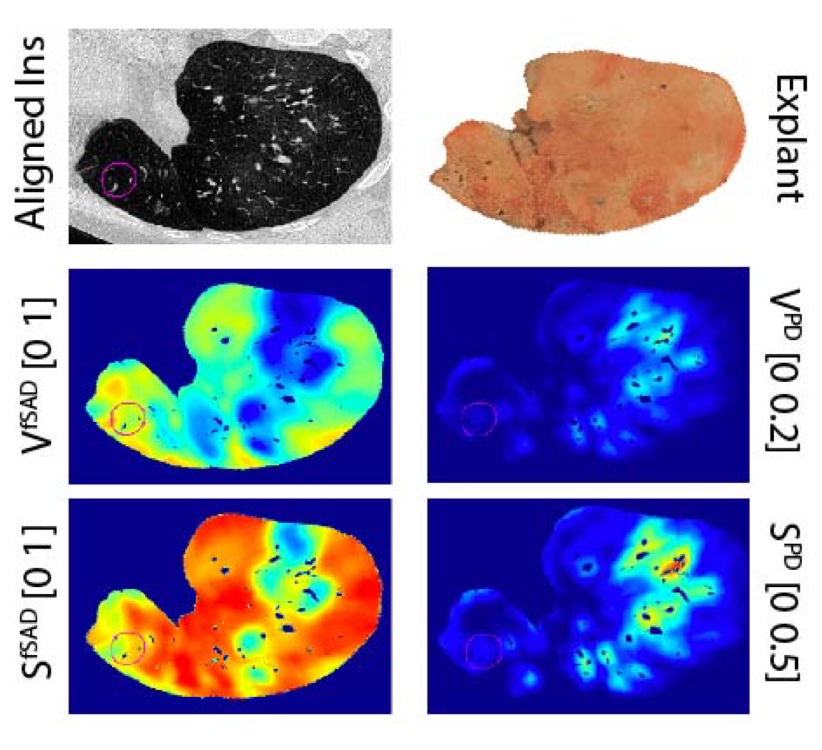
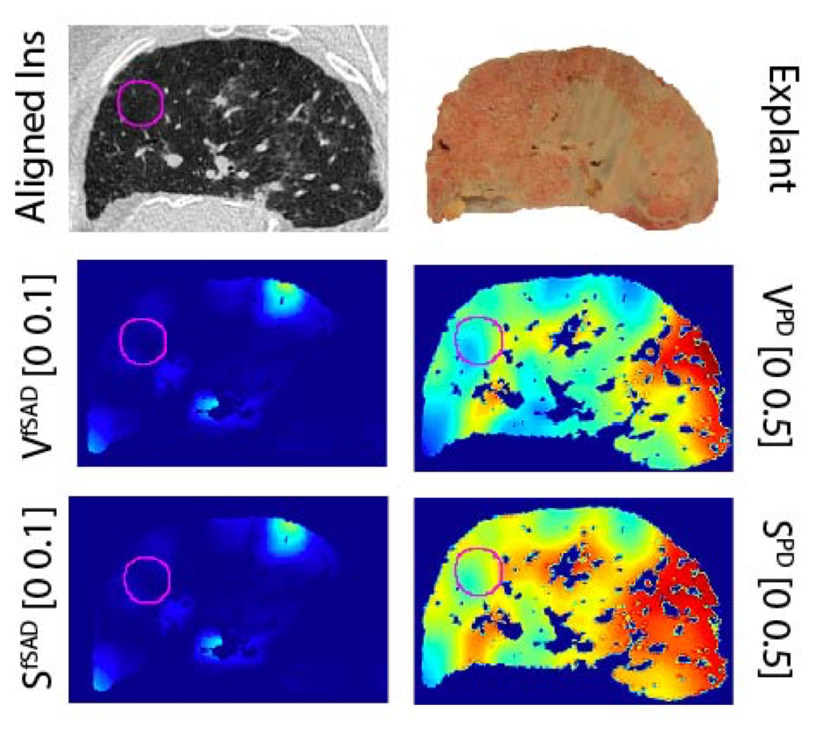
Chronic lung allograft dysfunction limits long-term survival following lung transplantation. Two distinct clinical phenotypes have been identified (bronchiolitis obliterans syndrome, BOS and restrictive allograft syndrome, RAS). Computed tomography (CT) aids in differential diagnosis as BOS typically shows mosaic attenuation and air trapping and RAS persistent pleuroparenchymal infiltrates. We aimed to assess the association between small airway disease (SAD)/parenchymal disease (PD), quantified using our topological-based CT analytical technique, and the concomitant presence of inflammatory cells. Paired CT scans, at inspiration and expiration, were obtained from 6 BOS and 6 RAS patients. We applied our topological Parametric Response Mapping (tPRM) approach to the paired CT scans to generate surface area maps that represent the extent and distribution of SAD and PD within the lungs (S SAD and S PD, respectively). Higher and lower values of S represent diffuse and clustered disease, respectively. We then spatially aligned the in vivo S SAD and S PD to the inflated and fixed explant specimen at the moment of redo-transplantation or autopsy. The explant specimen was further sampled and 4 random samples from each lung underwent microCT and quantitative immunohistochemistry for B-cells, CD4, CD8, neutrophils, eosinophils, mast cells and macrophages. BOS samples showed a higher S SAD compared to RAS (0.52 vs 0.13, P<0.0001), while RAS patients showed elevated S PD (0.30 vs 0.15, P=0.01). Within the BOS samples, there was an association between S SAD and the volume fraction of neutrophils (R=0.43, P=0.04) and macrophages (R=0.40 and P=0.05). Within RAS, the same association between S SAD and neutrophils was observed (R=0.49, P=0.02), as well as an association with the number of terminal bronchioles identified on microCT (R=-0.52 and P=0.01). For parenchymal disease, we could not find an association between inflammatory cells and S SAD in BOS. In contrast, we observed an inverse association between the volume fraction of neutrophils and S PD in the RAS group (R=-0.63, P=0.0015). Small airway disease quantified using our tPRM approach to analyzing paired CT scans is strongly associated with a neutrophilic inflammatory response in both phenotypes of CLAD, suggesting a renewed interest in the neutrophil as culprit in CLAD.

Read Our Paper

Research Icon
Research Icon