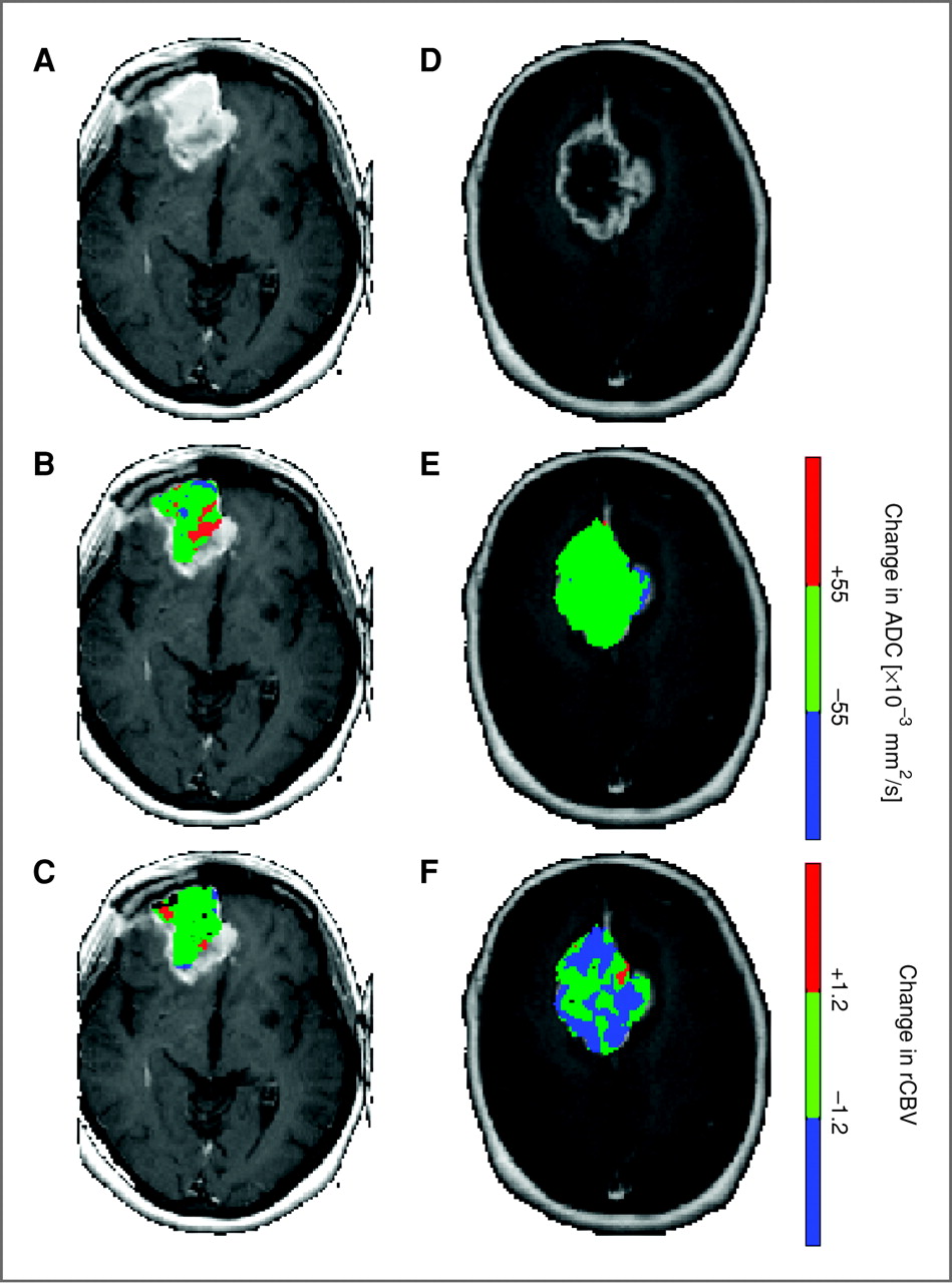
Imaging biomarkers capable of providing early cancer treatment response assessment would allow the opportunity to individualize patient care. For this purpose, images can be obtained which detect treatment-associated alterations in tissue properties including cellular viability, vascular function and volume, and biochemical and molecular responses. In this study, multiparametric quantification of perfusion- and diffusion-weighted MRI brain tumor maps was accomplished using a novel voxel-by-voxel analysis approach known as the parametric response map (PRM). The PRM composite imaging biomarker was found to provide early identification of patients resistant to standard chemoradiation. Validation of the PRM imaging biomarker would allow for routine clinical application as an early identifier of patients who may benefit from alternative treatment strategies.
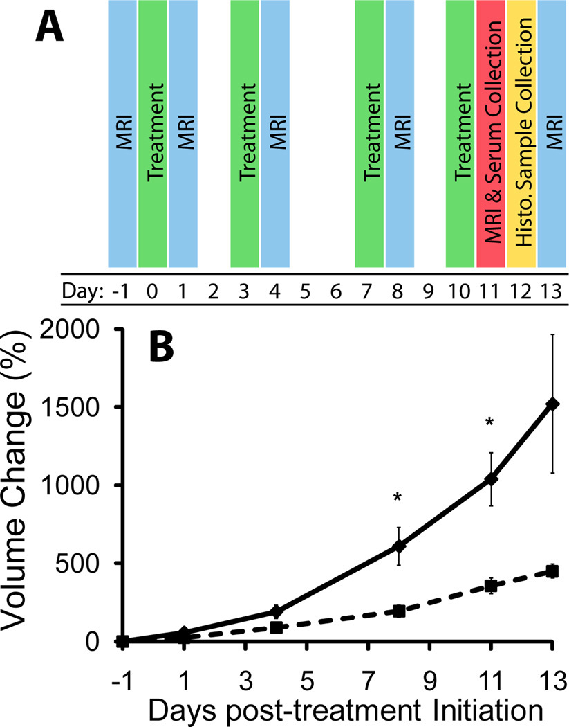
Read Our PaperVascular-targeted therapies have shown promise as adjuvant cancer treatment. As these agents undergo clinical evaluation, sensitive imaging biomarkers are need to assess drug target interaction and treatment response. In this study, dynamic contrast enhanced MRI (DCE-MRI) and diffusion-weighted MRI (DW-MRI) were evaluated for detecting response of intracerebral 9L gliosarcomas to the antivascular agent VEGF-Trap, a fusion protein designed to bind all forms of Vascular Endothelial Growth Factor-A (VEGF-A) and Placental Growth Factor (PGF). Rats with 9L tumors were treated twice weekly for two weeks with vehicle or VEGF-Trap. DCE- and DW-MRI were performed one day prior to treatment initiation and one day following each administered dose. Kinetic parameters (Ktrans: volume transfer constant, kep: efflux rate constant from extravascular/extracellular space to plasma, and vp: blood plasma volume fraction) and the apparent diffusion coefficient (ADC) over the tumor volumes were compared between groups. A significant decrease in kinetic parameters was observed 24 hours following the first dose of VEGF-Trapin treated versus control animals (p<0.05) and was accompanied by a decline in ADC values. In addition to the significant hemodynamic effect, VEGF-Trap treated animals exhibited significantly longer tumor doubling times (p<0.05) compared to the controls. Histological findings were found to support imaging response metrics. In conclusion, kinetic MRI parameters and change in ADC have been found to serve as sensitive and early biomarkers of VEGF-Trapanti-vascular targeted therapy.
Read Our Paper