RESULTS: Angular Kinematics
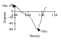
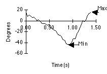
Segment angle. Motion Analyze was
used to calculate the shank angle over the complete duration of one
gait cycle. The shank rotated counterclockwise for most of the gait
cycle. Clockwise rotation of the shank occurred during the swing
phase from toe off to heel strike. Counterclockwise rotation occurred
from heel strike to mid-stance. The range of motion for our control
subject was 80 degrees, while the range of motion for our subject
with MS was 60 degrees. Maximum angle for both subjects was 20
degrees. The minimum angle of our control subject was -60 degrees,
and the minimum angle for the subject with MS was -40 degrees.
|
|
|
|
Figure 3. Shank angle in the normal walk (left) and the
MS walk (right). Shank ankle is determined from the angle
between the shank segment and the vertical axis. Anatomical
position corresponds to an angle of 0 degrees, full
extension. A positive slope of the curve represents
clockwise rotations of the shank while a negative slope
represents counterclockwise shank rotation.
|
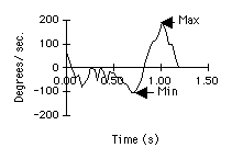
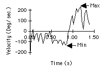
Segment velocity. Shank velocities
were similar for able-bodied and MS subjects. The maximum angular
velocity occurred during the swing phase for both subjects. The
maximum angular velocity of the shank for the control subject was 180
degrees/sec, while the maximum angular velocity for the subject with
MS was 200 degrees/sec. The maximum velocity for the control subject
occurred at 1 second. The maximum velocity for the subject with MS
occurred at 1.2 second. The minimum angular velocity of the shank for
the control subject was -100 degrees/sec and the minimum angular
velocity of the shank for the MS subject was -110 degrees/ sec.
|
|
|
|
Figure 4. Shank angular velocity in the normal walk
(left) and the MS walk (right). Positive velocity represents
clockwise rotation of the shank.
|
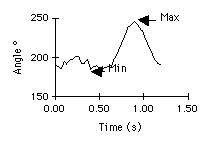
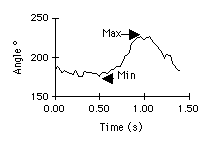
Joint angle1. The maximum knee joint
angle for the control subject was 250 degrees, and the maximum angle
for the subject with MS was 225 degrees. The maximum knee joint angle
occurs during the swing phase of the gait cycle as the knee is the
most flexed. The minimum knee joint angle for both subjects was 180
degrees. This minimum angle occurs during heel strike as the knee is
most extended. The range of motion at the knee for the normal subject
was 70 degrees and the range of motion at the knee for the subject
with MS was 45 degrees.
|
|
|
|
Figure 5. Knee joint angles in the normal walk (left) and
the MS walk (right). Knee joint angle is calculated as the
obtuse angle between the thigh segment and the shank
segment. 180 degrees corresponds to anatomical position.
Knee flexion occurs when the knee angle is greater than 180
degrees.
|
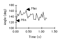
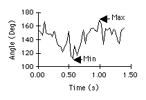
Joint angle 2. The maximum ankle
joint angle for the control subject was 160 degrees, while the
maximum angle for the subject with MS 170 degrees. The maximum ankle
angle occurs with maximum dorsiflexion, which occurs during heel
strike of the gait cycle. The minimum ankle angle for the control
subject was 125 degrees, and the minimum angle for the MS subject was
110 degrees. The minimum ankle angle corresponds to maximum plantar
flexion, which occurs during toe off of the gait cycle. The
able-bodied subject had a range of motion about the ankle joint of 35
degrees. The subject with MS had a range of motion of 60 degrees. The
minimum angle occurred at 0.80 sec for the control subject, and the
minimum angle for the MS subject occurred at 0.50 sec.
|
|
|
|
Figure 6. Ankle joint angles in the normal walk (left)
and the MS walk (right). Ankle joint is calculated
counterclockwise from the shank segment. Anatomical position
for the ankle joint is 90 degrees.
|
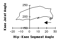
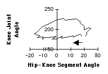
Angle-Angle Plot. The graph of
knee angle versus thigh angle represents the combined actions of the
knee flexion-extension and thigh forward-backward rotation. The
coordination of the thigh and knee was similar for able-bodied and MS
subjects. After toe off, the thigh rotates forward about the hip
joint and the knee flexes to its minimum angle. After the minimum
angle, the knee extends until just before foot strike while the thigh
rotates forward. During stance the thigh rotates backwards, and the
knee flexes then extends.
|
|
|
|
Figure 7. Coordination of the knee joint angle and the
thigh segment angle in the normal walk (left) and the MS
walk (right). Arrow points towards the direction of walk.
The curve is followed in a counterclockwise direction.
|


