METHODS
The subject in this analysis is a twenty-one year-old female college student. She is 5'5" and weighs 130 pounds. She is wearing a one-piece bathing suit in both motions. In the first motion she is barefoot and in the second motion she is wearing full foot swimming flippers in size seven. There are markers placed in four anatomical positions. The shoulder is marked at the humeral head. The hip is marked at the greater trochanter. The knee is marked at the joint line between the femur and tibia. The ankle is marked at the lateral malleolus.
|
|
|
|
Figure 1. Joint markers in barefoot walking (left) and flipper walking (right). Joint markers were placed on the shoulder, hip, knee, and ankle. | |
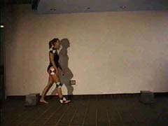
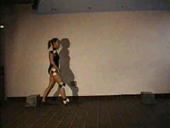
The data was collected in front of a white wall on a brown tile floor. The motions were filmed using a Sony high-8 video camera that was placed facing the subject's right side in order to film the gait. The room was light overhead and an additional spotlight was used directly on the subject. The subject was instructed to walk normally from marker to marker while barefoot and then while in flippers. The trials chosen for analysis best captured a full stride between the two markers with the best clarity.
The videotaped images were digitized at 30 frames/s using FusionRecorder on Macintosh computers in the New Media Center at the University of Michigan. The digital video files were trimmed using MoviePlayer so that the data files contained only the frames between the start and end of the movements. A custom utility (QT->PICT) was used to convert the QuickTime movie files into a series of individual frame files in PICT format for use with the Motion Plus software. Shoulder, hip, knee, and ankle joint markers were digitized using Motion Capture. Joint marker coordinate data were exported in spreadsheet format to Excel for biomechanical analysis using MotionAnalyse.