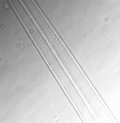
Amphipleura pellucida(Kütz.) Kütz.

                                                                                                                    
Length: 113

Width: 7

Striae: 40


Collection 1141a

Other images

More information


Return to Home Page Return to Genera List Return to Species List


Authority information


Kützing, F. 1844. Bacill., p. 103, pl. 3, fig. 32


Collection information


1141 Plankton, outside Muskegon north pier, Sec. 29, R17W, T10N, Muskegon County, Michigan. Diverse flora. Coll: E. F. Stoermer, 25 June 1966.


Other images


This is a bright field image.


This is a DIF image.


This is a bright field image of another specimen.


This is an enlargement of the end of the valve.


This is an image of the striae taken with oblique lighting.


More information


Long considered an excellent specimen for testing microscope objectives, Amphipleura pellucidais one of a few natural specimens to be employed as such. The microscope was, for a time, the most important scientific instrument and development of its capabilities was the object of much research. Resolving power, the ability to determine detail clearly, was a constant driving force. Diatoms were used as test specimens and the literature of that period has many references as to which objective could resolve what specimen(s) and the detail that particular objective could resolve of the specimen(s). Test plates of diatoms were used to evaluate the resolving power of objectives. Amphipleura pellucidawas the final test specimen on many test plates because of the fine structure of the cell wall. Stoermer and Pankratz (1964) give a concise summary of the historical work in reference to wall structure.
Being able to resolve the striae of Amphipleura pellucidademonstrates the capability (resolving power) of an objective and the level of skill by the microscopist. In an investigation of the effects of wave length of light and mounting media on the ability of microscope objectives to resolve the dots comprising the stria Trivelli and Lincke 1932 used Amphipleura pellucida as their test specimen. Stoermer (1996) utilized Amphipelura pellucida as a test specimen in a demonstration of the application of image analysis to diatom identification.

  • Ecology
    Cox (1975) emphasizes Amphipleura pellucidaas a free-living organism. Stoermer et al. (1965) state that Amphipleura pellucidain cultures grows in tubes. Andresen (personal experence) has collected Amphipleura pellucidain inland lakes in gelatineous masses but did not examine the mass for tubes.
    Stoermer (1980) states the disrtibution of Amphipleurataxa in the Laurentian Great Lakes is somewhat unusual. Amphileura pellucidais very widly distributed, mainly found in benthic associations and may get entrained into the plankton in disturbed areas where it may reach significant numbers. High populations of Amphipleura pellucidaare characteristic of the Green Bay water mass and can be found in the plankton of other more eutrophic regions. Lowe (1974) characterizes Amphipleura pellucidaas alkaliphilous, found in waters of pH range 6.2-8. He states it is a cosmopolitan, hard water taxon. Stoermer and Yang (1970) have Amphipleura pellucidaoccurring in low numbers in many samples as it gets injected into the plankton from its primarily benthic habitat. It is characteristic of disturbed habitats and always a minor constituent of the assemblage (Stoermer and Ladewski 1976). Based on field data it has its absolute abundance at 13°C and appears to be more commonly found in the fall.
             
         
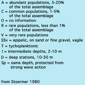
    A. arctica V R R   O V O   O O O   SSv T I-D
    A. pellucida C C A   C C A   R R C   SSv T Sp-I


    Size ranges and morphology


  • Length = 80-140 µm
  • Width = 7-9 µm
  • Striae = 40/10 µm
  • Other morphological features

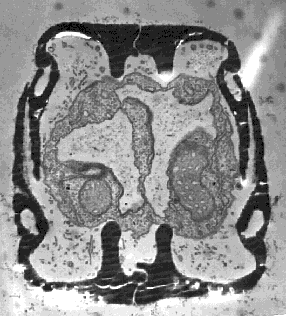
    This is an unpublished transmission electron micrograph taken by E. F. Stoermer showing a cross section through a cell at the end. Visible here are the silica ribs which form the area where the raphe is present. Notice that these project into the cell and are not external structures. Cytoplasmic contents, mitochondria and cytoplasm are visible. Notice also the chambers in the side walls (mantle) of each valve.
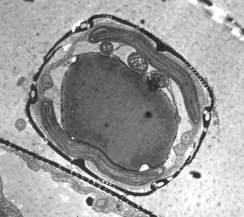
    This is an unpublished transmission electron micrograph taken by E. F. Stoermer showing a cross section through the middle of a cell. Visible here are plastids, mitochondria and a lipid droplet. The chambers in the side walls (mantle) are present suggesting these structures run the length of the cell.