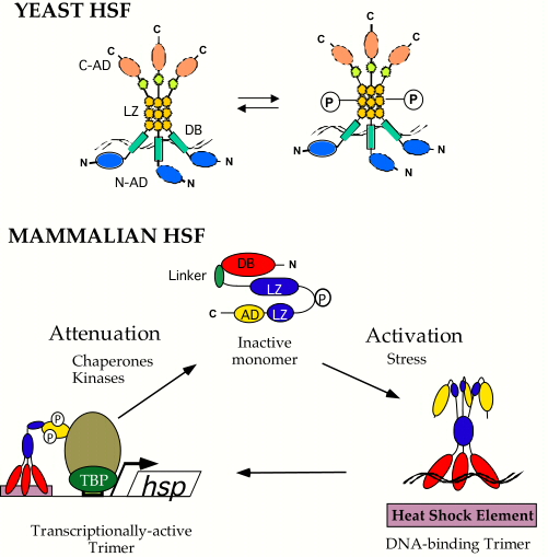
| My colleagues and I are interested in how organisms sense and respond to specific physiological and environmental changes. One area focuses on the mechanisms by which heat shock protein (Hsp) expression is induced in response to stress, pathophysiological conditions, pharmacological agents and normal cell growth and differentiation. Hsps are a class of highly conserved proteins that function in protein folding, assembly, targeting, processing and degradation and, as such, are often elevated in their levels in response to the stresses associated with growth and disease. Heat Shock Factors (HSFs) are a family of highly conserved transcription factors that respond to stressful conditions to activate the expression of Hsp genes. As shown in the figure to the right, the bakers yeast HSF exists in cells largely as a DNA bound homotrimer which, in response to stress, is hyperphosphorylated and transcriptionally active. Although yeast HSF is activated by multiple, distinct stresses, our results suggest that different signal transduction pathways and protein kinases communicate with HSF in response to disctinct stresses. We are interested in delineating these pathways to understand how cells use a single transcription factor to respond to multiple stresses. Humans and mice have multiple distinct HSF isoforms that appear to respond to unique signals and activate the expression of distinct target genes. As shown in the figure to the right, under non-stressful conditions human HSF1 (hHSF1) exists largely as a monomer unable to bind DNA with high affinity, which is thought to be sequestered through intramolecular interactions across coiled-coil domains, through its linker domain, and through inter-molecular interactions with some of the Hsps themselves. In response to stessful conditions, hHSF1 is thought to form homotrimers which bind to Hsp gene promoters, is hyperphosphorylated and transcriptionally competent. We have developed a powerful yeast-based genetic system to dissect out many of the important details for hHSF1 regulation, to identify novel proteins that regulate hHSF1 and to understand how distinct mammalian HSF isoforms activate different target genes. To learn more about our studies on Heat Shock Factors, please see some of our recent publications. |
| Another interest focuses on how cells regulate the acquisition and distribution of the trace metal copper (Cu). Cu is essential for a wide variety of enzymatic activities and biological processes including respiration, iron mobilization, neuropeptide modification and connective tissue maturation. Furthermore, several disease states are associated with abnormalities in Cu homeostasis. We have cloned genes and cDNAs encoding plasma membrane high affinity Cu transporters from bakers yeast and fission yeast, and from mice and humans. Given the power of yeast genetics, and the similarity between processes in yeast and humans, we are investigating the structure, function and transcriptional regulation of yeast Cu transporters. Yeast cells lacking these Cu transporters exhibit enzymatic and phenotypic properties that reflect deficiencies in Cu-dependent enzymes, suggesting that mammals defective in Cu transporters may present with abnormal neurological development, anemia and other symptoms . Interestingly, the yeast Ctr1 Cu transporter is regulated both at the level of abundance, by a Cu sensing transcription factor and at the level of protein trafficking, by Cu stimulated endocytosis. Furthermore, we have identified highly conserved domains within the Cu transport family members and are investigating their structure and function in Cu uptake and in Cu-stimulated changes in protein localization. These studies are carried out through biochemical, genetic, cell biology and molecular biology using the yeasts S. cerevisiae and S. pombe in flies and through the generation of mice with targeted deletions of Cu transporter genes. The figure to the right and the legend below summarize what is known about Cu homeostasis in yeast cells. The figure below shows the structural similarities of high affinity Cu transporters from yeast to mammals. To learn more about our work in Cu, please see some of our recent publications. | Copper homeostasis in S.cerevisiae
|
Model for Cu homeostasis in Saccharomyces cerevisiae. Work in a number of laboratories has allowed us to formulate a model of Cu homeostasis in yeast cells. Under Cu starvation conditions, Cu is transported into yeast cells with high affinity, following reduction from Cu(II) to Cu(I) by the Fre plasma membrane Cu(II)/Fe(III) ion reductases. The high affinity Cu transporters Ctr1 and Ctr3 mediate the passage of Cu across the plasma membrane, with mobilization of Cu to cytochrome c oxidase (Cyt Ox) by Cox17 (and possibly Sco1), to cytosolic superoxide dismutase (Cu/Zn Sod1) by CCS, and to a P-type ATPase, Ccc2, by Atx1, respectively. Once Cu reaches Ccc2, it is transported to the lumen of the Golgi/endosome compartment. Here, four Cu atoms are assembled with Fet3, with the assistance of the Gef1 chloride channel, which provides Cl- ions that allosterically facilitate Cu loading onto Fet3. The Fet3-Ftr1 high affinity Fe-transport complex assembles at the plasma membrane, resulting in the formation of an active Fe transport complex in which Fet3 is the multi-Cu ferroxidase and Ftr1 is the Fe permease subunit. Furthermore, when cells are grown during Cu scarcity, the Cu-sensing transcription factor Mac1 activates expression of high affinity Cu-uptake genes including CTR1, CTR3, and FRE1/7, while repressing Fe transporter gene (such as FET3) expression. The asterisks indicate yeast Cu homeostasis proteins for which homologs have been identified in humans and mice.
![]() |
Structural comparisons of yeast and mammalian high affinity Cu transporters. High affinity Cu ion transporters have been isolated from the yeasts S. cerevisiae and S. pombe, and from mouse and human. These transporters are highly structurally similar. The putative Cu-binding motif Met-X2-Met-X-Met is found eight, five, and two times in Ctr1 (Saccharomyces cerevisiae), Ctr4 (Schizosaccharomyces pombe), and hCtr1 and mCtr1(human and mouse), respectively (depicted by the vertical ovals). The predicted transmembrane spanning domains (TM) are depicted by the dark ovals. |
home | research interests | lab members | alumni | publications | lab links